Close Menu
OziDent
    Facebook X (Twitter) Instagram
    Latest Post
    • What Truly Motivates Dentists? 8 Key Driving Forces Behind the Grin
    • From Burnout to Balance: Discovering Ikigai in the Life of a Dentist
    • Adult ADHD Dentist
    • Marketing Strategies for Endodontists!
    • 7 Proven Ways to Gain New Dental Patients in the UAE
    • INBDE Exam Content Breakdown
    • NBDE I & II Vs INBDE
    • The Five Stages of Dental Career
    Facebook X (Twitter) Instagram
    OziDent
    • Home
    • Articles
      • Education & Licensing
    • Contact Us
      • Authors
      • F.A.Q.
      • Privacy Policy
      • Affiliations
      • Site Map
      • Our Principles
    • Send Your C.V.
    • Register / Login
    • Dr.Ozaibi د.العزيبي
    • Dental Buys
    OziDent

    Cysts of the Jaw – Odontogenic Cysts

    0
    By Mohammed AlShanbari on September 3, 2013 Dental Education, Featured

    Odontogenic Cysts

    In this article, we will write about the different types of odontogenic cysts, its special features, diagnosis  and proper management and treatment. To read about the other Type of cysts of the Jaw:
    1. Cysts of the Jaw – Nonodontogenic Cysts
    2. Cysts of the jaw – Pseudocyst
    F15.large_

    Radicular cyst

    Other Names

    Periapical cyst, apical periodontal cyst, or dental cyst.
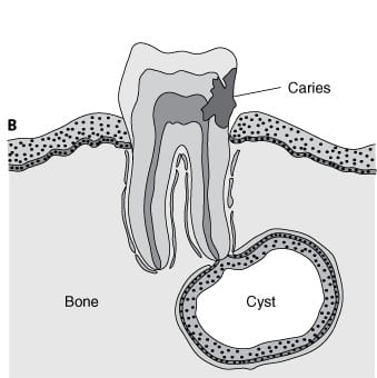
    Radicular cyst
    Radicular cyst

    Features

    • Most common.
    • Inflammatory cyst because in the majority of cases it is a consequence to pulpal necrosis with an associated periapical inflammatory response. ( Rests of Malassez )
    • Related to the apex of non vital tooth or related to lateral canal.
    • cyst formation occurs as a result of epithelial proliferation, which helps to separate the inflammatory stimulus ( necrotic pulp ) from surrounding bone.

    Clinical features

    • Asymptomatic, unless associated with infection or intra-cystic pressure.
    • Swelling of the jaw is the chief complaint.
    • Soft fluctuant swelling, bluish in color beneath the mucosa.
    • Cause bone resorption.
    • On palpation may feel bony and hard unless if bone resorption occurs thus it feels soft and rubbery.
    • Slight Male Prevalence.
    • Occurring in 3rd to 6th decade.
    •  60% in Maxilla Canine or incisors.

    Radiographically

    • Location apex of a non-vital tooth, but occasionally on the mesial or distal of the teeth.
    • Cannot be differentiated from periapical granuloma.
    • Radiolucency ( round or ovoid ) with narrow, opaque margin that is continuous with the lamina Dura.
    • well­ defined cortical border.
    • Root Resorption of offending tooth and occasionally  the adjacent tooth.
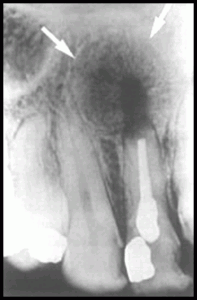
    Radicular Cyst Healing After Endodontic Treatment, New Bone Is Growing Towards The Center From Periphery
    Radicular Cyst Healing After Endodontic Treatment, New Bone Is Growing Towards The Center From Periphery
    Well Defined Cortical Boundary Of Radicular Cyst
    Well Defined Cortical Boundary Of Radicular Cyst

    Differential Diagnosis

    • Odontogenic keratocyst.
    • Stafne’s developmental salivary gland defect.


    Treatment

    • Extraction of the associated non-vital tooth and curettage of the apical zone.
    • RCT may be performed with an apicoectomy ( direct curettage of the lesion ).
    • The most often used is only RCT, since most periapical lesions are granulomas and resolve after removal of the inflammatory stimulus ( necrotic pulp ).
    • Surgery ( apicoectomy and curettage ) is done for persist lesions, presence of a cyst or inadequate RCT. Residual cyst may occur if the cyst lining incompletely removed.


    Residual cyst

    Features

    • It arises either due to improper surgical elimination of a radicular cyst or extraction of the related tooth without removal of the cyst

    Clinical Features

    • Asymptomatic and often is discovered on radiographic examination of an edentulous area.
    • Its clinical and histological characteristics are identical to those of a radicular cyst
    • Radiographically it will seen as a radiolucency of variable size at the site of a previous tooth extraction.


    Radiographically

    • Often in the mandible and always above the inferior alveolar nerve canal.
    • Periphery and shape. cortical margin shape is oval or circular.


    Differential Diagnosis

    • Odontogenic keratocyst.
    • Stafne’s developmental salivary gland defect.


    Treatment

    • complete removal of the cystic lesion ( enucleation ).
    •  In large cyst; Marsupialization is achieved by creating a window to evacuate the cyst and to decrease the intracystic pressure.  Refer to Article to explain the Difference between.


    Lateral periodontal cyst

     Features

    • Unkeratinized developmental cyst.

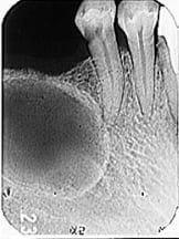
      Lateral Periodontal Cyst In The Mandibular Region
      Lateral Periodontal Cyst In The Mandibular Region
    • Usually related to a vital tooth.
    • Proliferation of rests of dental lamina.
    • It is rare, mostly seen in males over 50 years of age.
    • Gingival cysts in adult are histogenetically and pathologically similar.

    Clinical Features

    • Asymptomatic.
    • Mandibular premolars and bicuspid region and occasionally in the incisors areas.
    • Maxillary lateral incisors region.
    • 4th and 5th decades of life most effected.

    Radiographically

    • Well-delineated, round or teardrop-shaped.
    • Unilocular radiolucency with opaque margin along the lateral surface of a vital tooth root.


    Diffrentail Diagnosis

    • A small odontogenic Keratocyst.
    • Small mental foramen.
    • small neurofibroma
    • a radicular cyst at the foramen of a lateral (accessory) pulp canal.

    Treatment

    • Local excision is generally curative.
    • In case of multilocular ( botryoid ) cyst seems to have recurrence potential, follow up, therefore suggested for treated multilocular odontogenic cyst.


    Gingival cyst ( Newly Born)

    Features

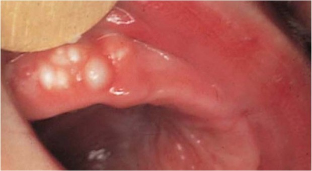
    • It is a cyst that arises from the rest cells of the dental lamina
    • Solitary nodules on the crest of the alveolar ridge of the new born or very young infants. it appears as a multiple, small, firm, white or grayish-white nodules on the alveolar ridge .
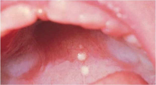
    • Epstein´s pearls are cystic nodules found on the hard palate or at the junction of hard and soft palate.

    Treatment

    • No treatment is necessary, because spontaneous rupture usually occur early.


    Gingival cyst ( Adults)

    Features
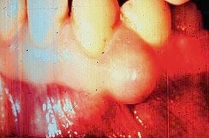
    Gingival cyst
    Gingival cyst
    • A small developmental cyst of the gingival tissues derived from the remnants of dental lamina. It appears as a small circumscribed painless swelling, same color as the color of the gingiva.

    Treatment

    • Local excision.


    Epstein’s pearls 

    Epstein's pearls are keratin-filled cystic lesions lined with stratified squamous epithelium. They appear as small, whitish lesions along the midpalatine raphe and contain no mucous glands
    Epstein’s pearls are keratin-filled cystic lesions lined with stratified squamous epithelium. They appear as small, whitish lesions along the midpalatine raphe and contain no mucous glands

    Bohn’s nodules

    Bohn's nodules are mucous gland cysts, often found on the buccal or lingual aspects of the alveolar ridges and occasionally on the palate. They are multiple, firm, and grayish white in appearance. Histologically they show mucous glands and ducts
    Bohn’s nodules are mucous gland cysts, often found on the buccal or lingual aspects of the alveolar ridges and occasionally on the palate. They are multiple, firm, and grayish white in appearance. Histologically they show mucous glands and ducts

    Dental lamina

    Dental lamina cysts are found only on the crest of the alveolar mucosa. Histologically, these lesions are different because they are formed by remnants of dental lamina epithelium. They may be larger, more lucent, and fluctuant than Epstein pearls or Bohn nodules and are more likely to occur singly.
    Dental lamina cysts are found only on the crest of the alveolar mucosa. Histologically, these lesions are different because they are formed by remnants of dental lamina epithelium. They may be larger, more lucent, and fluctuant than Epstein pearls or Bohn nodules and are more likely to occur singly.

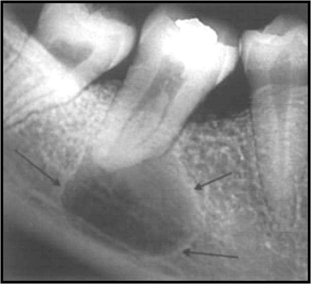
    Dentigerous or follicular cyst

    Definitions
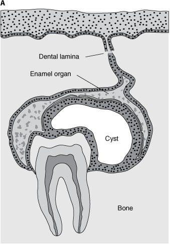
    Dentigerous Cyst
    Dentigerous Cyst

    Dentigerous Cyst is one which encloses part or all of an unerupted crown of a tooth. It is attached to the junction where the tooth enamel meets the root (amelocemental junction) and arises in the follicular tissues covering the fully formed crown of the unerupted tooth.

    Features

    • Second most common.
    • Arise from reduced enamel epithelium of the dental follicle of an unerupted tooth.
    • Developmental.
    • Attached to the tooth cervix ( ECJ ).
    •  Enclose the crown of unerupted tooth.

    Clinical Features

    • Associated with 3rd molar and maxillary canine; mostly IMPACTED.
    • More in Mandible.
    • 2nd to 3rd decades.
    • More in Males

    Radiographically

    • Well-defined, unilocular or occasionally multilocular Radiolucency.
    • The unerupted tooth is often displaced.
    • n the mandible the RL may extend to  the ramus or to the body of mandible.
    • In maxillary canine region the cyst may extend into the sinus or to the orbital floor.
    • Resorption of adjacent tooth root maybe seen.

    Treatment

    • Extraction of the tooth and enucleation.
    • Marsupilization of the cyst to allow for decompression and subsequent shrinkage of the lesion.

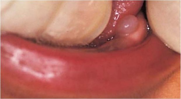
    Eruption cyst

    Definition

    The eruption cyst is the soft tissue analogue of the follicular cyst.
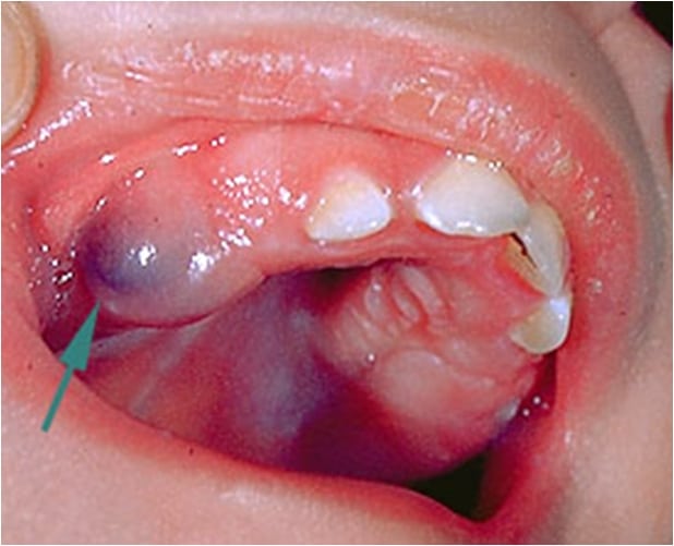
    Eruption cyst
    Eruption cyst

    Features

    • Developmental.
    • Result from fluid accumulation within the follicular space of an erupting tooth.
    • The epithelium lining this space is simply reduced.


    Clinical Features

    • It appears as soft fluctuant blue to dark red mass on the alveolar ridge in place of any erupting tooth, particularly molar and canines.


    Radiographically

    • Negative as it is soft tissue cyst above the crown of unerupted tooth.

    Treatment

    • No treatment is needed, because the tooth erupts through the lesion, the cyst disappears spontaneously without complication.

    Odontogenic Keratocyst (Primordial Cyst)

    Features

    • Arises from reduced enamel epithelium, dental lamina rests and malassez rests.
    • May exhibit aggressive clinical behavior.
    • Associated with nevoid basal cell carcinoma.
    • It can be found any where in the jaw.
    • Highly recurrence after initial surgical intervention.
    • characterized by keratinizatoin and budding cyst lining.
    • Has a wide age range but there is a peak incidence in the 2nd and 3rd decade with a smaller peak in the 5th decade.
    • The cysts are more common in males and 70% occur in the wisdom tooth and ramus region of the lower jaw.
    • Can Reach Large sizes without causing expansion, because it predominantly enlarges anterior-posterior direction.
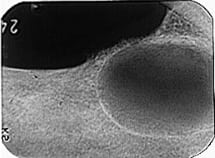
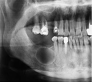
    Odontogenic Keratocyst
    Odontogenic Keratocyst
    Odontogenic Keratocyst
    Odontogenic Keratocyst
    Odontogenic Keratocyst
    Odontogenic Keratocyst


    Clinical Features

    • Arises from reduced enamel epithelium, dental lamina rests and malassez rests.
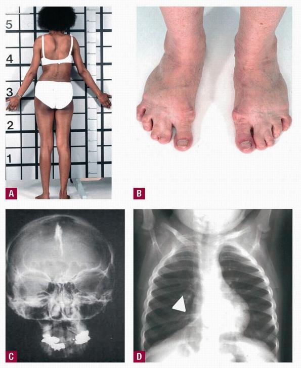
      Gorlin syndrome Manifestations
      Gorlin syndrome Manifestations
    • May exhibit aggressive clinical behavior.
    • Associated with nevoid basal cell carcinoma. (Gorlin syndrome) it has many manifestations:
      • Oral – multiple odontogenic keratocysts, cleft lip or palate.
      • Skin – multiple nevoid basal cell carcinoma
      • Skeletal – rib anomalies, vertebral deformities, polydactyly (Birth defect characterized by the presence of more than the normal number of fingers or toes)
      • Central nervous system – calcified falx cerebri, brain tumors
    • It can be found any where in the jaw.
    • Highly recurrence after initial surgical intervention.
    • Characterized by keratinizatoin and budding cyst lining.


    Radiographically

    • Well-circumscribed Radiolucency with smooth radiopaque margins.
    • Multilocular most common in large lesions.
    • It may cause buccal expansion.
    • It may cause displacement of roots or IAC.
    • Resorption of the root is common.
    • May be associated with missing or unerupted tooth.
    •  It maybe difficult to distinguish from Dentigerous cyst.
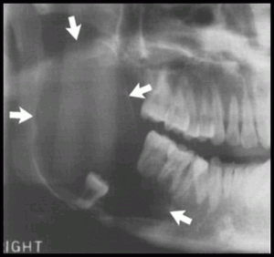
      Odontogenic Keratocyst
      Odontogenic Keratocyst

     Differential Diagnosis

    • Dentigerous cyst
      • The cyst is likely to be an OKC if the cyst is connected to the tooth at a point apical to the cementoenameljunction or if no expansion of the cortical plates has occurred.
    • Ameloblastoma.
    • Odontogenic myxoma.
    • A simple bone cyst.

    Treatment

    • Surgical excision with peripheral osseous curettage or osteoectomy is the preferred method of management.
    • The use of chemical cauterization of the cyst in Highly recurrence cyst has been advocated.
    • In Large cyst; Marsupilization, followed by enucleation, may be an attractive alternative.

    Reasons for recurrences

    • Thin and fragile cystic epithelium.
    • Has satellite cysts (daughter cysts).
    • Epithelial lining has intrinsic growth potential.
    • Has increased mitotic activity.
    • Patients with nevoid basal cell syndrome have particularly tendency for recurrence of keratocysts.


    Glandular Odontogentic Cyst

    Features

    • Rare.
    • Developmental odontogenic cyst.
    • Most common site of occurrence is the front region of the lower jaw.
    • They present as slow-growing, painless swellings.
    • The cyst has a potentially aggressive, locally invasive nature.
    • Recurrence tendency.
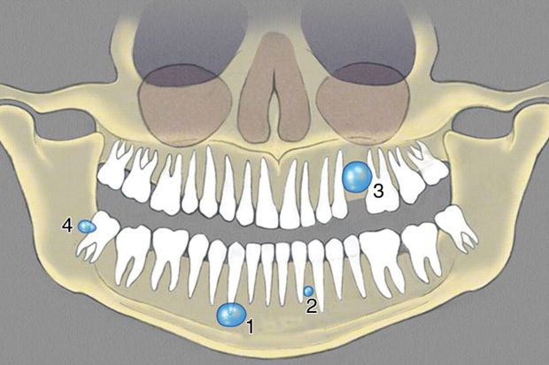
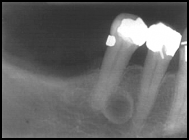
    1 = Apical Radicular Cyst, 2 = Lateral Radicular Cyst, 3 = Residual Radicular Cyst, 4 = Paradental Cyst
    1 = Apical Radicular Cyst, 2 = Lateral Radicular Cyst, 3 = Residual Radicular Cyst, 4 = Paradental Cyst

    Paradental Cyst

    Features

    • Arises along partly erupted wisdom teeth that is involved by pericoronitis.
    • Mostly in the lower jaw.
    • Located at the cheeks side of the tooth


    Sources

    • Ibn Sina University Curriculum By Dr. M. Shanbari.
    • INTELLIGENT DENTAL  RADIOGRAPHIC APPEARANCE OF CYSTS PART 2 by Dr. MEIFONG.
    • Wikipedia.


    OziDent Members Only

    The rest of article is viewable only to site members,Please Register and/ or Confirm registration via EmailHere.
    If you are an existing user, please login.
    Existing Users Log In

    Prove your humanity


    Forgot password? Click here to reset
    New User? Click here to register
    Share. Facebook Twitter Pinterest LinkedIn Tumblr Email
    Avatar photo
    Mohammed AlShanbari
    • Website

    Dentist and Guest Author on OziDent. He earned his BDS in 2011 from Ibn Sina College, Jeddah, Saudi Arabia with honors. Currently working as a Demonstrator in Umm al-Qura University, Makkah, Saudi Arabia. His Goal is to be a Maxillofacial surgeon. In His Free time he enjoys Photography and Trying different Cuisine.

    Related Posts

    From Burnout to Balance: Discovering Ikigai in the Life of a Dentist

    Adult ADHD Dentist

    Marketing Strategies for Endodontists!

    Comments are closed.

    Login Status

    Prove your humanity


    Forgot?  Register
    CV Creating for Dentists
    Join Our Study Group
    National Dental Board Exam Study Group
    Recent Posts
    May 4, 2025

    What Truly Motivates Dentists? 8 Key Driving Forces Behind the Grin

    April 18, 2025

    From Burnout to Balance: Discovering Ikigai in the Life of a Dentist

    September 14, 2024

    Adult ADHD Dentist

    May 29, 2024

    Marketing Strategies for Endodontists!

    May 6, 2024

    7 Proven Ways to Gain New Dental Patients in the UAE

    December 12, 2023

    INBDE Exam Content Breakdown

    Login Status

    Prove your humanity


    Forgot?  Register
    Help Support Ozident
    About OziDent

    We are a Group of enthusiastic dentists and dental specialist who share a common goal of giving back to the public.

    Top Posts
    • What Truly Motivates Dentists? 8 Key Driving Forces Behind the Grin
    • From Burnout to Balance: Discovering Ikigai in the Life of a Dentist
    • Adult ADHD Dentist
    Instagram
    علشان عدم وضوح ورش العمل … جمعتها ورتبتها من اجل تسهيل الحضور @sharjahef #sef2025 
———————————————
سعادتكم غايتنا و رضاكم نيتنا 
وصحتكم هدفنا وتعليقاتكم تهمنا
———————————————
للتواصل:
‏‎‏Whatsapp: 0562861010
‏‎‏Snap: @dr.ozaibi
‏Email: mohsen@ozident.com
‏‎‏‎ابوظبي - Abu Dhabi, UAE
‏________ د. محسن العزيبي __________
دكتور محسن سعيد سيف العزيبي
- طبيب أسنان عام.
- علاج العصب بدون الم.
- خبرة طويلة في علاج الأطفال واستخدام الغاز الضاحك للاطفال.
- مدرب حياة معتمد دولياً.
- حاصل علي ماجستير تسويق و تجارة
- مؤسس الموقع التعليمي لطب الاسنان OziDent.com
‏________ D R . M O H S E N __________
‏‎‏Dr. Mohsen said Saif Al Ozaibi
‏- General Dentist
‏- Painless Root canal 
‏- child management under nitrous oxide sedation. 
‏- Certificated Life coach 
‏- An MBA holder in marketing
‏- founder of the dental educational website OziDent.com
____________________________________
وما توفيقي الا بالله واختموا إعجابكم بـ ( ماشاء الله )
دقة المواعيد مهمة لذلك 
علشان اعطيكم حقكم راعوا موعدكم 
____________________________________
التسويق و التصميم تحت رعاية شركة OziTect 
‏www.ozitect.com | mail@ozitect.com | @ozitect
____________________________________
‏#explorer #اكسبلور
    Follow on Instagram

    All Rights Reserved to the respected Sources, OziDent 2012 - 2023 | Created and Designed by OziTect.com | Protected and Managed by DMCA.com Protection Status

    Type above and press Enter to search. Press Esc to cancel.

    Manage Cookie Consent
    We use technologies like cookies to store and/or access device information. We do this to improve browsing experience and to show (non-) personalized ads. Consenting to these technologies will allow us to process data such as browsing behavior or unique IDs on this site. Not consenting or withdrawing consent, may adversely affect certain features and functions.
    Functional Always active
    The technical storage or access is strictly necessary for the legitimate purpose of enabling the use of a specific service explicitly requested by the subscriber or user, or for the sole purpose of carrying out the transmission of a communication over an electronic communications network.
    Preferences
    The technical storage or access is necessary for the legitimate purpose of storing preferences that are not requested by the subscriber or user.
    Statistics
    The technical storage or access that is used exclusively for statistical purposes. The technical storage or access that is used exclusively for anonymous statistical purposes. Without a subpoena, voluntary compliance on the part of your Internet Service Provider, or additional records from a third party, information stored or retrieved for this purpose alone cannot usually be used to identify you.
    Marketing
    The technical storage or access is required to create user profiles to send advertising, or to track the user on a website or across several websites for similar marketing purposes.
    Manage options Manage services Manage {vendor_count} vendors Read more about these purposes
    View preferences
    {title} {title} {title}