

In this Article, we will describe and state a differential diagnosis for the most common Periapical radiolucent lesions that means that it is located in the periapical area and appears radiolucent. When we say non-pulpal origins it means the tooth is vital (most cases) and the teeth are mostly not effected (unless its periodontitis or malignant tumor) but the source of the lesion isn’t due to the pulp.
Case Questions
Here are some Case questions :D… try to express the differential diagnosis (at least 3).
- A RL Periapical cyst found in the mid line of the anterior part of the maxilla , adjacent teeth are vital but displaced, the palate is expanded. (Answers At the end of the Article)
- A RL Periapical lesion in 33 year old African, located near the apex of the lower central inscisor the PDL is intact and there is no effect on the teeth.
Periapical Radiolucent Lesions of Non-Pulpal Origins
Definitions
- Periapical: comes from the words peri: around and apical:root.
- RL: Radiolucent: it express that the region/area/object didn’t absorb the radiation but it’s transparent/translucent thus more radiation reaches the sensor/film leading to a darker area. (Low mineral content: Tissue other than bone).
- RO: Radiopaque: it express that the region/area/object absorbed the radiation thus less radiation reaches the sensor/film leading to a lighter area. (High Mineral Content: Bones and teeth).
- Unilucular: From the words uni: means one and lucular means lobes, it means the lesion appears as one mass.
- Monolucular: From the Words Mono: means multiple. it means the lesions appears as multiple mass in one lesion area.
- Corticated: means the lesion contains an outer cortex thus it appears with radiopaque outer boarder.
[divider scroll_text=””]
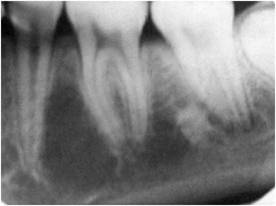
Cementoma First Stage
Another Name: PCD (Periapical Cemento-osseous dysplasia).
Age: Middle Age.
Ethnicity: African.
Site: Apex of multiple lower Incisors.
Description: Round, Monolucular and often multiple.
Periodontium: Periodontal ligament space is intact.
Dentition: Teeth are Vital, No displacement, No resorption.
Features:
- Small ( up to 1 cm in diameter).
- Shape:
- Early Stage: Radiolucent and not corticated.
- Intermediate Stage: Radioopaque with radioluceney around the Apical region
- Late Stage: Dense radioopaque lesion with a thin Radiolucent line.
Differential Diagnosis:
- Traumatic Bone cyst.
- Radicular Cyst.
- Periapical Granuloma.

Periodontitis
PCD: Periapilcal Cemento-osseous dysplasia.
Description: Localized.
Dentition: Severe destruction around the effected roots of the teeth ( An Endo-Perio Lesion).
Differential Diagnosis:
- Radicular cyst.
- Periapical Granuloma.
- Eosinophilic Granuloma
- Traumatic Bone cyst.

Traumatic Solitary Bone Cyst
Age:Young under 20.
Site: Mandible at the premolar/molar area..
Description: Monolucular, Moderately well corticated and Moderately Well Defined.
Periodontium: Scalloping of the cyst wall between the adjacent teeth.
Dentition: Adjacent teeth are Vital, NO Displacement and No resorption.
Features:
- Bone Cavity not lined with epithelium.
- Unknown etiology.
- Asymptomatic.
- Scalloping (Superior margin of the cyst squeezes between the roots of the adjacent teeth.
Differential Diagnosis:
- Radicular cyst.
- PCD 1st stage.
- Central Giant Cell Granuloma. (CGCG)
- Odontogenic Keratocyst
Nasopalatine Duct cyst
Site: Midline, Anterior Maxilla.
Description: Round or oval, Unilocular, Corticated (unless infected) and Well defined
Periodontium: May cause Palatal Expansion..
Dentition: Adjacent teeth are Vital, Displaced and “Rarely resorbed”..
Features:
- Most Common Non-Odontogenic Cyst.
- Raise from the Epithelial remnants of the duct or incisive canal.
Differential Diagnosis:
- Large incisive Foramen
- Radicular Cyst


Eosinophilic Granuloma
Age: Adolescent and young adults..
Site: In Jaws (75% in mandible).
Description: Round, Monolocular, Not Corticated and it could be either Localized or multiple.
Periodontium: PDL destruction “Loose teeth”
Dentition: No Effect on teeth (Vital).
Features:
- Benign Lytic Lesion of bone characterized by proliferation of Langerhans cell.
Differential Diagnosis:
- Periodontitis.
- Radicular Cyst.
- Squamous Cell Carcinoma.
- ill defined metastatic tumor.
- Malignant Salivary Gland Tumor.

Malignant tumor Periapical Radiolucent lesions
Features:
Malignant tumor which are pericapical monolucular RL:
- Metastatic Tumor.
- Squmaos Cell Carcinoma
- Fibrosarcoma.
- Chondorsarcoma.
- Osteosarcoma
Differential Diagnosis:
- Ameloblastoma.
- Giant Cell Granuloma.
- Ossifying Fibroma.
- Aneurismal Cyst.
- Benign Non-odontogenic Tumor.
Answers
- Answer Question 1: naso-palatine duct cyst, large incisive foramen, Periodontitis.
- Answer Question 2: Trumatic bone cyst, Cementoma 1st stage, Radicular cyst.
OziDent Members Only
The rest of article is viewable only to site members,Please Register and/ or Confirm registration via EmailHere.If you are an existing user, please login.


