In this new section on OziDent, We provide a quick dental guide for all General Dentists to manage what might seem at first a complex case but with the correct information and knowledge should be easy mange and handle. In this article we tackle the the different causes of Maxillary Canine Impaction, Proper diagnosis, prognosis, possible treatments.
Maxillary Canine Impaction
Facts
- The 2nd most common impacted tooth.
- Twice as common in females.
- Twice as common in the maxilla than mandible.
- 8% of impacted canines are bilateral.
- 1/3 is labial located impaction while 2/3 are palatally located impactions.
Etiology
The main reason for impaction of maxillary canine isn’t really understood but studies showed that labially impacted canines were due to insufficient arch space, while the palatal is due to genetic or Guidance theory. Guidance theory states that distal surface of lateral incisor’s root is the guidance line used by the canine to erupt and any disturbance (absence, malformed…etc) to this surface will eventually cause an impaction.
Sequelae
This is the squelae of problems that will occur if the impacted canine (in its different positions) is left alone, it could cause one of theses problems or a combination of them:
- Malpositioning of the impacted tooth.
- Migration of the neighbouring teeth.
- Loss of arch length.
- Internal resorption and External root resorption of the impacted tooth and neighbouring teeth,
- Dentigerous cyst formation.
- Infection (with partial eruption)
- Referred pain and combinations of the above sequelae.
[divider scroll_text=”SCROLL_TEXT”]
Diagnosis
Diagnosis is based on both clinical and radiographic examination:
Clinical Examination
- Delayed eruption of the permanent canine or delayed shedding of the deciduous canine.
- Absence of a normal labial canine bulge
- Presence of a palatal bulge.
- Delayed eruption, distal tipping, or migration of the lateral incisor.
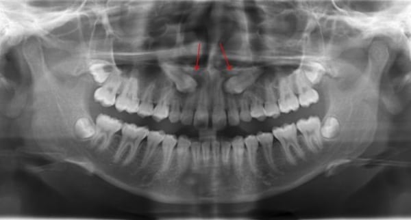
Radiographic Examination
- Depending on the provided equipment a panorama X-ray is sufficient to identify the impaction type and prognosis. While if the panorama isn’t readily available 2 peri-apical X-rays in two different directions will be enough to guide you to the proper diagnosis.
[divider scroll_text=”SCROLL_TEXT”] Treatment
GP Interceptive Treatment
Depending on the X-ray, the GP has a few solutions at his finger tips. These factors are:
- Has the Canine passed the midline of the Lateral Incisor.
- Is the Deciduous canine still present.
- There is sufficient space for the permeant canine to erupt.
Here are a Few possible Scenarios A General dentist might face after diagnosing the an impacted canine.
- Deciduous Canine is still present but mobile, X-ray shows that the Permeant canine hasn’t passed the distal side of the lateral incisor, there is no root resorption, Sufficient arch space for canine . In this case, leave the case as it will normalise its self or if the child is a bit old extract the Deciduous canine.
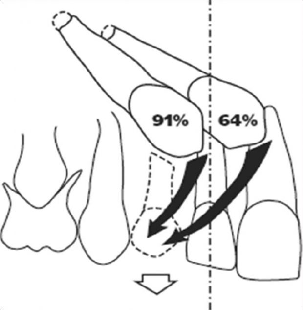
- Deciduous Canine is still present, X-ray shows that the Permeant canine Has passed the distal side of the lateral incisor but not the mid line of the lateral, there is no root resorption, Sufficient arch space for canine . In this case, extract the Deciduous canine. ( success rate is 91%, According to Williams)
- Deciduous Canine is still present, X-ray shows that the Permeant canine Has passed the distal side and the mid line of the lateral incisor, there is no root resorption, Sufficient arch space for canine . In this case, extract the Deciduous canine. ( success rate is 64%, According to Williams).
- No sufficient space for the canine or Impacted canine has passed the mesial side of the lateral incisor , some signs of root resorption, patient age is above 10. In these possible situations, Please refer to an orthodontist immediately.
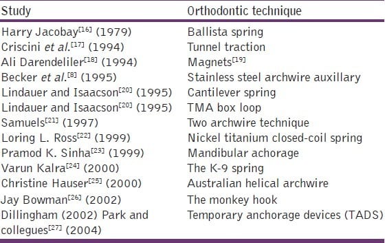
Orthodontic Treatment
here is a list of Orthodontic techniques used to manage and treat impacted canine, Because this article is targeted towards General Dentists, I will not go into details.
[divider scroll_text=”SCROLL_TEXT”] Summery
Impacted Maxillary canine, even with the latest technologies is still considered a complex yet not impossible matter to handle, it depends most importantly on early diagnosis by the dentist to give it the best prognosis and treatment, proper dialogue between the genral dentist and orthodontist and a positive attitude. Remember General Dentist to always check a child 10 years of age of the following indicators:
- Extreme Lateral Tilting.
success rate 91% if extraction is done before the canine passes the midline of the lateral and 64% if after. - Deciduous Canine , is still fixed and show no signs of movement.
- No normal Labial bulge or presence of an abnormal palatal bulge.
If you see one or more of these indicators, PLEASE DO AN X-RAY, to evaluate the case, because if you intercept it early by a simple extraction you can save the patient so much time, effort and money.
[divider scroll_text=”SCROLL_TEXT”]
Sources
- J Pharm Bioallied Sci. 2012 August; 4(Suppl 2): S234–S238. Link.
- Ranjit Manne, 2012, Dental science: Impacted canines: Etiology, diagnosis, and orthodontic management. Link.
- Crystallakeoralsurgery.com
OziDent Members Only
The rest of article is viewable only to site members,Please Register and/ or Confirm registration via EmailHere.If you are an existing user, please login.